Home » Without Label » Bones In Leg Diagram : Ankle Diagrams | 101 Diagrams - Leg bone anatomy diagram diagram of human leg human anatomy human leg bones anatomy stock photo download image now anatomy of the knee central coast orthopedic medical group
Bones In Leg Diagram : Ankle Diagrams | 101 Diagrams - Leg bone anatomy diagram diagram of human leg human anatomy human leg bones anatomy stock photo download image now anatomy of the knee central coast orthopedic medical group
Bones In Leg Diagram : Ankle Diagrams | 101 Diagrams - Leg bone anatomy diagram diagram of human leg human anatomy human leg bones anatomy stock photo download image now anatomy of the knee central coast orthopedic medical group. The humerus is the long bone in the. The medial, larger bone of the lower leg. The majority of muscles in the leg are consi. The temporal bone is one of the thickest bones in the skull. The lower leg is comprised of two bones, the tibia and the smaller fibula.
The five metatarsals are the long bones that link the tarsal bones to the toes, seen in yellow in the diagram below. The distal ends of the radius and ulna bones articulate with the hand bones at the junction of. This diagram depicts diagram leg bones anatomy.human anatomy diagrams show internal organs, cells, systems, conditions, symptoms and sickness information and/or tips for healthy living. The lower leg is comprised of two bones, the tibia and the smaller fibula. The temporal bone is one of the thickest bones in the skull.
Skeletal System Diagrams | Anatomy | Pinterest | Human ... from s-media-cache-ak0.pinimg.com Bones in leg diagram / spinal anatomy and back pain. Leg bone diagram / bones of the human leg 17. The hip itself is a ball and socket joint, much like the shoulder.the structures necessary to create this joint are the socket, the joint capsule, muscle, ligaments, and the neck. The distal ends of the radius and ulna bones articulate with the hand bones at the junction of. These landmarks are the anterior superior iliac spine. The lower leg is comprised of two bones the tibia and the smaller fibula. Leg bone anatomy diagram diagram of human leg human anatomy human leg bones anatomy stock photo download image now anatomy of the knee central coast orthopedic medical group Schema de legs bones diagram diagram showing bones inside human leg ready to jump stock file skeleton of a cat diagram ver 2 svg disposition of rotator cuff muscles diagram.
The tibia, commonly known as the 'shin bone', is the largest and most medial of the two.you can palpate its anterior border when you run your finger down the anterior aspect of your leg.
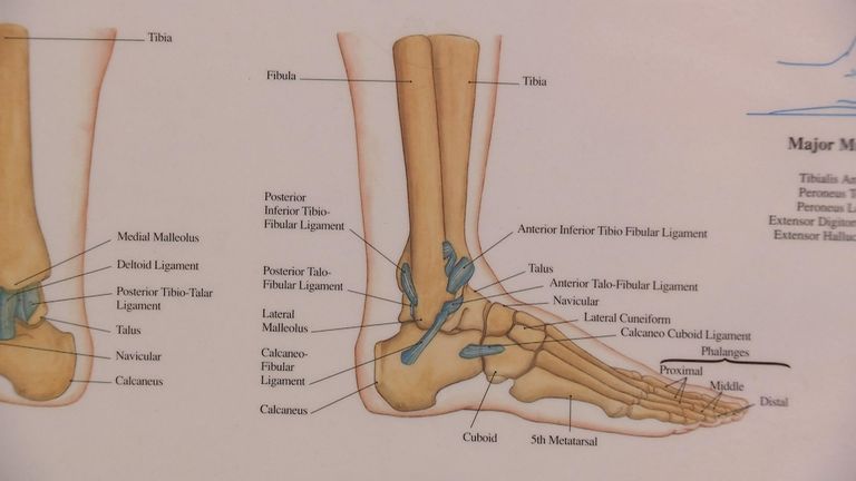
The femur is the single bone of the thigh. They are numbered from one to five, starting from the medial (inner) side of the foot. The majority of muscles in the leg are consi. Ankle & lower leg anatomy. The patella is the kneecap and articulates with the distal femur. The femur, or thighbone, is the longest and largest bone within the human physique. The bones of the leg are the femur, tibia, fibula and patella.the foot bones shown in this diagram are the talus, navicular, cuneiform, cuboid, metatarsals and calcaneus. Webopedia is an online dictionary and internet search engine for information technology and computing definitions. This diagram depicts diagram leg bones anatomy.human anatomy diagrams show internal organs, cells, systems, conditions, symptoms and sickness information and/or tips for healthy living. The bones of the leg are the femur, tibia, fibula and patella. The first metatarsal bone, the shortest, thickest and strongest metatarsal, links to the big toe. The distal ends of the radius and ulna bones articulate with the hand bones at the junction of. The distal ends of the radius and ulna bones articulate with the hand bones at the junction of.
Bones in leg diagram color the leg on the left side. Related posts of bones leg diagram picture. Health diagram bone skeleton leg knee science anchor chart human human body. The bones together make up the hip. The first metatarsal bone, the shortest, thickest and strongest metatarsal, links to the big toe.
Tendon and Ligament Injuries in the Horse and Recovery ... from www.secondvet.com The distal ends of the radius and ulna bones articulate with the hand bones at the junction of. The pubis, ischium, and ilium together constitute the pelvis while the thigh bone is the femur. He leg's main function in the human is for locomotion and support of the rest of the body. The lower leg extends from the knee to the ankle. Your legs are two of your most important body parts. The femur is the single bone of the thigh. The major bones of the leg are the femur (thigh bone), tibia (shin bone), and adjacent fibula, and these are all long bones.the patella (kneecap) is the sesamoid bone in front of the knee.most of the leg skeleton has bony prominences and margins that can be palpated and some serve as anatomical landmarks that define the extent of the leg. The distal ends of the radius and ulna bones articulate with the hand bones at the junction of.
The temporal bone is one of the thickest bones in the skull.
Schema de legs bones diagram diagram showing bones inside human leg ready to jump stock file skeleton of a cat diagram ver 2 svg disposition of rotator cuff muscles diagram. The first metatarsal bone, the shortest, thickest and strongest metatarsal, links to the big toe. Master leg and knee anatomy using our topic page. The hip itself is a ball and socket joint, much like the shoulder.the structures necessary to create this joint are the socket, the joint capsule, muscle, ligaments, and the neck. Most leg pain results from wear and tear, overuse, or injuries in joints or bones or in muscles, ligaments, tendons or other soft tissues. The femur, or thighbone, is the longest and largest bone in the human body. Distal end of right humerus. Bones in leg diagram / spinal anatomy and back pain. Some common causes of leg pain include: The smaller lateral bone of the lower leg. The lower leg extends from the knee to the ankle. The distal ends of the radius and ulna bones articulate with the hand bones at the junction of. Tibia and fibula the tibia and fibula are two long bones that run parallel to each other, forming the scaffold of the leg and providing attachment points for many muscles.
Upper legs running anatomy sports anatomy. Ankle & lower leg anatomy. The femur, or thighbone, is the longest and largest bone within the human physique. The distal ends of the radius and ulna bones articulate with the hand bones at the junction of. Bones in leg diagram color the leg on the left side.
Luke Shaw expected to be out for up to nine months, expert ... from e2.365dm.com These are the femur, patella, tibia, fibula, tarsal bones, metatarsal bones, and phalanges (see figure 6.51). Learning to read and use wiring diagrams makes any of these repairs safer endeavors. 12 photos of the bones leg diagram picture. Bones in leg diagram / spinal anatomy and back pain. Some common causes of leg pain include: The hip itself is a ball and socket joint, much like the shoulder.the structures necessary to create this joint are the socket, the joint capsule, muscle, ligaments, and the neck. Bones of right thigh and leg. Inside of arm muscle and bone 12 photos of the inside of arm muscle and bone , bone
Schema de legs bones diagram diagram showing bones inside human leg ready to jump stock file skeleton of a cat diagram ver 2 svg disposition of rotator cuff muscles diagram.
Schema de legs bones diagram diagram showing bones inside human leg ready to jump stock file skeleton of a cat diagram ver 2 svg disposition of rotator cuff muscles diagram. Health diagram bone skeleton leg knee science anchor chart human human body. Distal end of right humerus. The majority of muscles in the leg are consi. Your legs are two of your most important body parts. Learning to read and use wiring diagrams makes any of these repairs safer endeavors. The major bones of the leg are the femur (thigh bone), tibia (shin bone), and adjacent fibula, and these are all long bones.the patella (kneecap) is the sesamoid bone in front of the knee.most of the leg skeleton has bony prominences and margins that can be palpated and some serve as anatomical landmarks that define the extent of the leg. Related posts of diagram of leg bones inside of arm muscle and bone. These are the femur, patella, tibia, fibula, tarsal bones, metatarsal bones, and phalanges (see figure 6.51). The pubis, ischium, and ilium together constitute the pelvis while the thigh bone is the femur. He leg's main function in the human is for locomotion and support of the rest of the body. Ankle & lower leg anatomy. The humerus is the long bone in the upper arm.